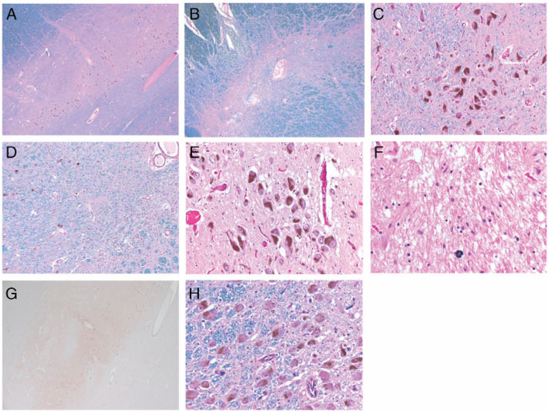
FIGURE 2:

(A–D) Photomicrographs of the left substantia nigra: rostral, medial levels in A and C; caudal in B; and caudal, medial levels in D. The loss of pigmented neurons is uneven and prevails at the caudal levels. Relatively sharp demarcation between foci without or with resilient neurons in D. (E–H) Photomicrographs of the caudal levels of the right substantia nigra in E–G and of the nucleus coeruleus in H. (E) The density of pigmented neurons is apparently normal within the medial third. (F) The loss is subtotal within the lateral third. (G) Neither Lewy body-containing neurons nor Lewy neurites were detected. (H) The neuronal density of the nucleus coeruleus is normal. Staining: Luxol Fast Blue counterstained with Hematoxylin and Eosin in A–D and H; Hematoxylin and Eosin in E and F; and a-synudein in G. Original magnification: ×25 in A, B, and G; ×100 in D; ×200 in C, E, and H; and ×400 in F.
