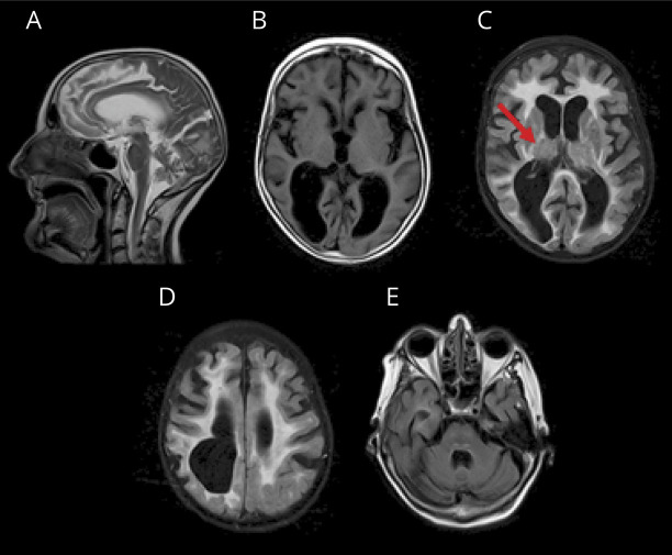
Figure 2. Mother's MRI.
(A) Thin corpus callosum, ventriculomegaly (atrophy), and cerebellar vermis atrophy on sagittal T2 image. (B) Axial T1 image shows ventriculomegaly and significant posterior white matter atrophy. Fluid-attenuated inversion recovery images reveal diffuse white matter hyperintensity (C, D) involving the posterior limb of the internal capsule (C, arrow) and corticospinal tracts and medial lemniscus in the pons (E).

