Figure 1.
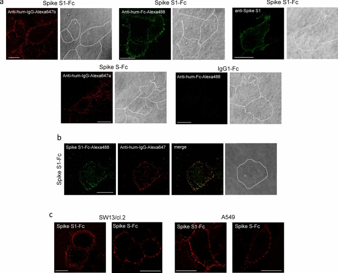
Detection of Spike protein constructs at the cell surface. (a) Vero cells plated on glass coverslips were incubated with recombinant Spike S1-Fc, Spike S-Fc, or IgG1-Fc, as indicated, for 1 h in the cold, as described in “Methods”, and the proteins retained on the cell surface were detected with the indicated antibodies. Anti-human (anti-hum) IgG-Alexa647a and Anti-human-IgG-Alexa647b are antibodies raised in goat and alpaca, respectively. (b) Cells were incubated with Spike S1-Fc labeled with Alexa488 (green) followed by anti-human IgG-Alexa647 (red) and the overlay of the two signals is shown (merge). In (a,b), bright field images in which the approximate cell contours have been highlighted are shown. (c) SW13/cl.2 or A549 cells were incubated with Spike S1-Fc or Spike S-Fc, which were detected by incubation with anti-human-IgG-Alexa647. Images shown are single confocal sections taken at mid-height of the cells. Bars, 20 μm.

