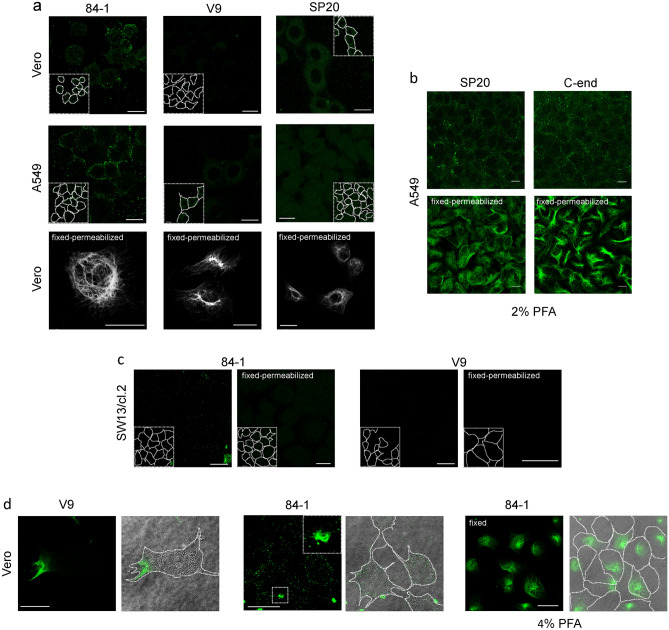
Figure 3.
Detection of vimentin in several cell types by several fixation and permeabilization protocols. When indicated, cells were fixed, or fixed and permeabilized, prior to immunodetection. (a) In the two upper rows, Vero or A549 cells were incubated with the indicated monoclonal primary antibodies at 1:200 dilution for 1 h in the cold, followed by incubation with Alexa-488-conjugated secondary antibodies at 1:200. At the end of the procedure cells were fixed with 4% (w/v) PFA in the cold. Representative images at mid-cell height are shown. In the lower row, Vero cells were fixed and permeabilized prior to detection of cytoplasmic vimentin with the same monoclonal antibodies. (b) A549 cells were incubated with the indicated anti-vimentin antibodies, and the corresponding secondary antibodies, before fixation (upper panels) or after fixation with 2% (w/v) PFA and permeabilization (lower panels) for detection of cytoplasmic vimentin. (c) Vimentin-deficient SW13/cl.2 cells, were incubated with the indicated anti-vimentin monoclonal antibodies and Alexa-488-conjugated anti-mouse immunoglobulins at 1:200, before fixation (left images) or after fixation and permeabilization (right images). (d) Vero cells were incubated with the indicated anti-vimentin antibodies as described above for detection of cell surface vimentin. Images shown illustrate representative cases of cells in which an area of the cytoplasm shows staining of filamentous vimentin (left panels), or that display fragments of vimentin filaments associated to their surface (middle panels). In the right panels, Vero cells were stained with anti-vimentin antibodies after fixation with 4% (w/v) PFA in the cold without performing any additional permeabilization step. In each case, images on the right show the overlay of the bright field and fluorescence images with the contours of the cells highlighted. Insets in (a,c) display the approximate cell contours drawn from bright field or overexposed images, whereas the inset in (d) middle panels shows a cell surface-associated vimentin bundle. Bars, 20 μm in (a,c,d) and 10 μm in (b).

