
Figure 4.
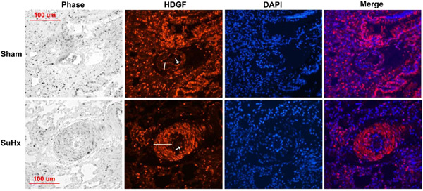
Hepatoma‐derived growth factor (HDGF) is highly expressed in Sugen/hypoxia (SuHx) rat pulmonary artery smooth muscle cells. The lung tissue architecture is shown in the phase panel and HDGF immunostaining (red florescent) with arrow indicating staining of the luminal endothelial layer and the bar the smooth muscle medial layer. Nuclei are stained blue (4′,6‐diamidino‐2‐phenylindole counterstaining) with merged images showing HDGF and nuclear colocalization. A 100 μM scale bar is included for reference