. 2020 Apr 29;10(28):16727–16731. doi: 10.1039/d0ra03291c

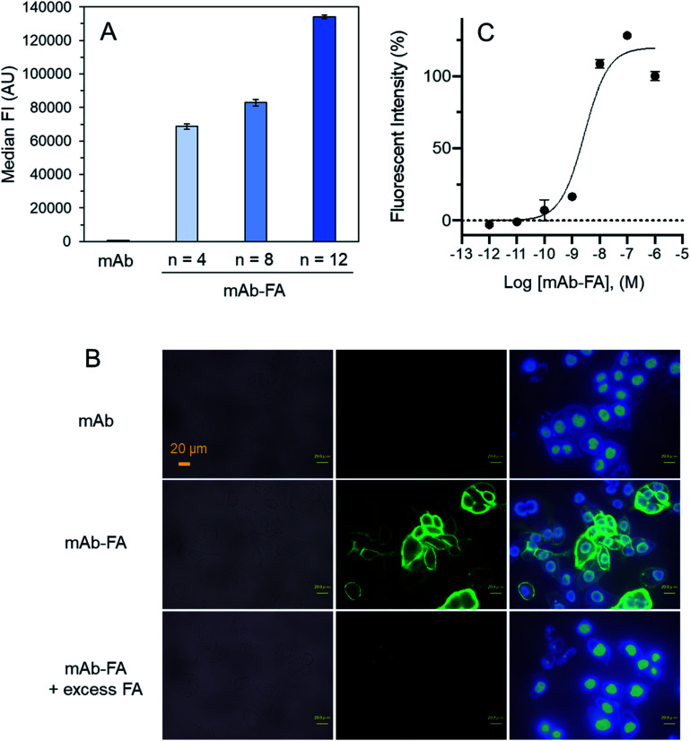
Fig. 4. mAb–FA binds to the FR-α expressed on IGROV-1 cells. (A) IGROV-1 cells were incubated with anti-CD20–FA conjugates with different linker lengths (PEG, n = 4, 8, 12). After a washing step, FITC-labelled anti-IgG Fc polyclonal antibody was added to each sample, and the median of fluorescent intensity (FI) was measured using flow cytometry. (n = 3, mean ± SEM.) (B) IGROV-1 cells were seeded and incubated overnight. mAb, mAb–FA, or mAb–FA + excess FA were added. After washing, mAb–FA bound to the cells was detected using anti-IgG-Fc polyclonal IgG. Cells were also stained with Hoechst 33342. (C) IGROV-1 cells were treated with increasing concentrations of mAb–FA, and the antibodies bound to the cells were quantified. (n = 3, mean ± SEM.).

Fig. 4