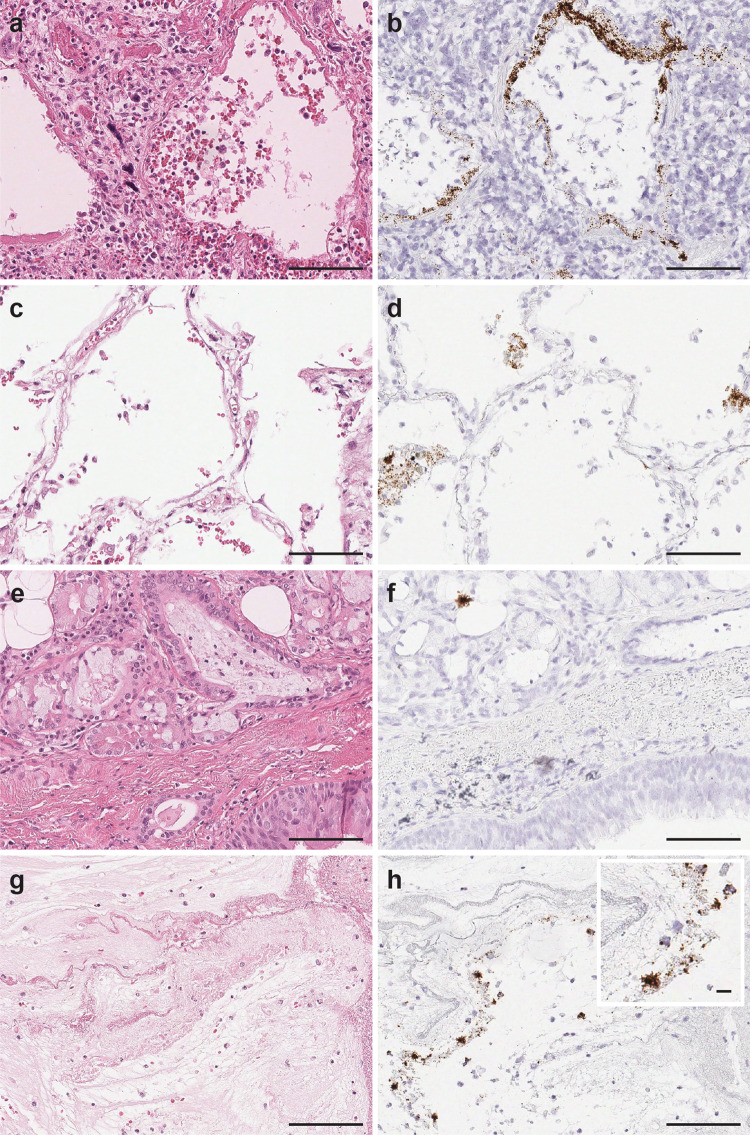
Fig. 1. SARS-CoV-2 virus detection in pulmonary parenchyma by SARS-CoV-2 spike RNA-ISH.
The viral signals were observed within the intra-alveolar hyaline membranes (a H&E and b SARS-CoV-2 spike RNA-ISH) and within the intact lining alveolar epithelial cells as well as in the desquamated alveolar epithelial cells (c H&E and d SARS-CoV-2 spike RNA-ISH). Viral particles were also noted in the nonalveolar region like bronchus as a cluster of viral signals within peri-bronchial sero-mucinous glands, as well as within the lining pseudostratified respiratory bronchial epithelium. (e H&E and f SARS-SoV-2 spike RNA-ISH). Viral signals were also detected in the necrotic and fibrinous material within damaged pulmonary tissue (g H&E and h SARS-CoV-2 spike RNA-ISH). Inset: Viral signals shown as individual punctate brown dots and clusters. Scale bars = 100 µm. Inset scale bar = 10 µm.

