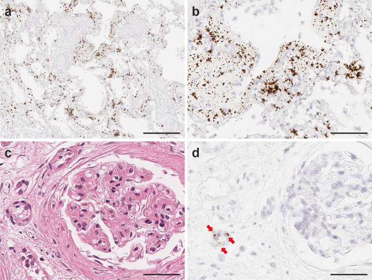
Fig. 6. SARS-CoV-2 infection in kidney transplant patient (patient 3) on immunosuppressant therapy.
Highly abundant viral signals were observed within the lung parenchyma exhibiting diffuse alveolar damage, reflecting the high SARS-CoV-2 virus infection load in Patient 3. The viral signals were detected in intra-alveolar hyaline membrane and interstitial fibroblastic proliferation region (a, b. SARS-CoV-2 RNA-ISH). Scattered viral signals were identified in the renal tubules (c H&E and d SARS-CoV-2 RNA-ISH). Scale bar = 500 µm in a, 100 µm in b, 50 µm in c, d.

