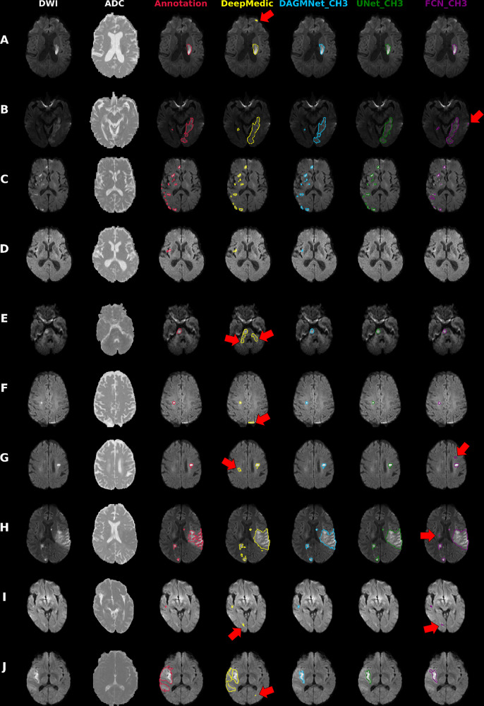
Fig. 7. Illustrative cases of models’ performances.
Columns from the left to the right: DWI, ADC, and overlays on DWI of the manual delineation (red), DeepMedic Predicts (yellow), DAGMNet_CH3 (our proposed model) predicts (blue), UNet_Ch3 (green), and FCN_CH3 (purple). A Typical lesion. B Case with inhomogeneity between DWI slices; note the high agreement of our proposed model with the manual annotation. C Multifocal lesions. D Small cortical lesion, detected exclusively by DeepMedic and our proposed attention model. E–G Typical false positives (arrows) of other models (DeepMedic in particular) in areas of: E “physiological" high DWI intensities; F DWI artifacts in tissue interfaces; in addition to the cortical areas, this was vastly observed in the basal brain, along the sinuses interfaces, and in the plexus choroids; G in possible chronic microvascular white matter lesions. H, I Cases in which the retrospective analysis favored the automated prediction, rather than the human evaluation for: H lesion delineation, and I lesion prediction (this case was initially categorized by evaluators as “not visible" lesion, but the small lesion predicted by our model was confirmed by follow-up). J Lesion of high-intense core but subtle boundary contrast, which ameliorates the discriminative power of all 3D networks. In this case, the patch-wise DeepMedic had the best agreement with manual annotation in the boundaries at the cost of detecting false positives (arrow).

