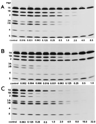
FIG. 2.
Fluorograms of PBPs of H. influenzae TK422 pretreated with AMP (A), CTX (B), and FRM (C). A 30-μl aliquot of each membrane fraction was incubated with 3 μl of each of the nonradioactive β-lactams diluted to various concentrations for 10 min and then further incubated with 3 μl of [3H]PEN for another 10 min. PBPs were visualized by autoradiography after exposure of the dried gels to X-OMAT film for 20 days at −80°C. The numbers identifying the lanes are concentration (in micrograms per milliliter).

