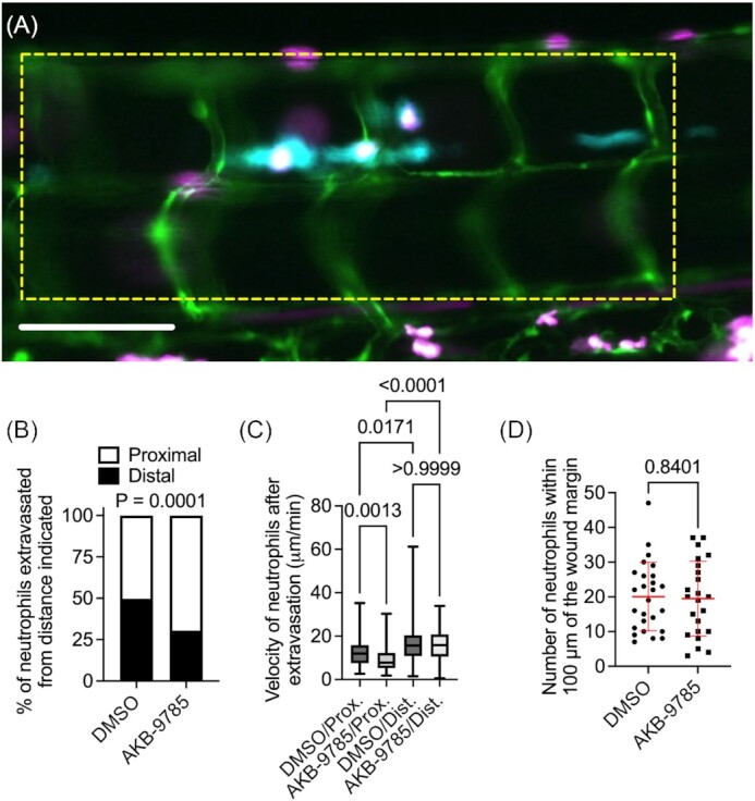
Figure 4.

Vascular normalization focuses neutrophil extravasation to blood vessels near granulomas in zebrafish embryos. (A) Representative image of a granuloma from a 4 dpi M. marinum-Cerulean (blue) infected Tg(Tg(kdrl:egfp, lyzC:dsred) s843, nz50 zebrafish embryo with pseudo coloring of blood vessels green and neutrophils magenta. Yellow dotted box outlines the two intersegmental vessel bounds around the central granulomas used to define proximal extravasation events. Scale bar represents 100 µm. (B) Quantification of neutrophil extravasation event source for neutrophils recruited to M. marinum granulomas, proximal defined as within 2 intersegmental vessels. Total number of events measured: DMSO = 153, AKB-9785 = 262; total embryos imaged: DMSO = 8, AKB-9785 = 12. (C) Quantification of neutrophil straight-line velocity after extravasation until neutrophil reaches M. marinum granuloma. (D) Counts of neutrophils within 100 µm of the tail amputation margin at 6 h postwounding. Each data point represents a single animal. Statistical test for (B) was performed by Fisher's exact test on raw counts, statistical test for (C) was performed by ANOVA with Dunn's multiple comparisons test, and statistical test for (D) was performed using unpaired T-test. (D) data are representative of two experimental replicates with similar numbers of animals.
