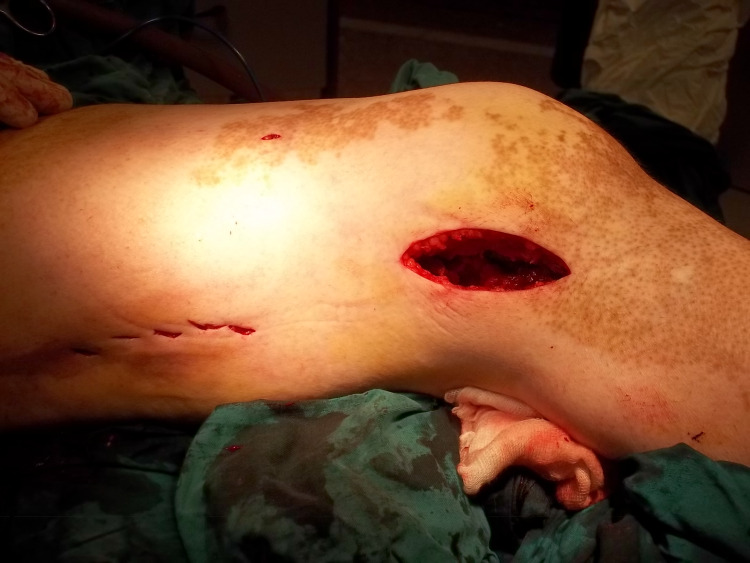
Figure 1. Lateral approach to the distal femur.
The patient is supine on the radiolucent table with a bump beneath the ipsilateral buttock. If there is no intra-articular extension, then a distal lateral incision is made starting just proximal to the lateral epicondyle and continuing distally. The incision is sharply continued through the iliotibial band and underlying vastus lateralis in line with their fibers. Dissection proceeds to bone without disruption of the overlying periosteum.

