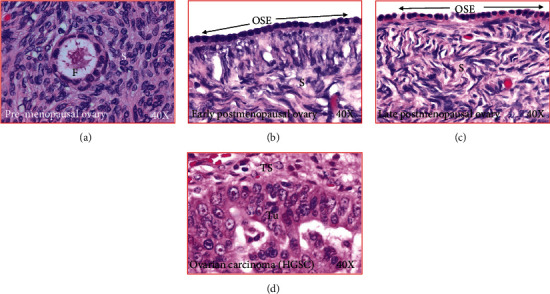
Figure 1.

Microscopic presentations of healthy ovaries and ovary with cancer. (a) Section of an ovary from a premenopausal subject. An embedded follicle is seen in the ovarian stroma. (b) Section of an ovary from a healthy early postmenopausal woman showing no embedded follicle in the stroma. The ovarian surface layer is seen to be composed of rounded or flat-type of epithelial cells. (c) Section of an ovary from a healthy late postmenopausal woman showing no embedded follicle in the stroma. The ovarian surface layer is seen to be composed of rounded or flat-type of epithelial cells. (d) Section of an ovarian high-grade serous carcinoma (HGSC) at late stage. OSE: ovarian surface epithelial layer; S: stroma; TS: tumor stroma; Tu: tumor; 40×: magnification.
