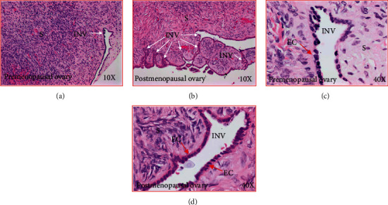
Figure 3.

Stromal invaginations (INV) in healthy ovaries in pre- and postmenopausal women. (a) Section of an ovary from a premenopausal woman showing an INV in the stroma at low magnification (10×). (b) Section of an ovary from a healthy woman at late postmenopausal stage (low magnification, 10×). Compared with premenopausal woman, many INVs of different sizes and shapes are seen in the ovarian stroma. (c) Section of an ovary from a healthy premenopausal woman (presented in (a)) showing an INV in the stroma at high magnification (40×). The INV consists of a single layer of rounded or occasionally tube-like epithelial cells. (d) Section of an ovary from a healthy late postmenopausal woman (presented in (b)) showing an INV in the stroma (at high magnification, 40×). As seen in the premenopausal ovary, the INV is consisted with a single layer of rounded or occasionally tube-like epithelial cells. EC: epithelial cells; S: stroma.
