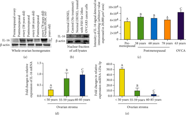
Figure 6.

Changes in IL-16 protein and gene expression in the ovary during aging. (a) Western blot showing IL-16 expression during ovarian aging. (a) Changes in IL-16 protein expression in the ovary during aging. A very weak or faint immunoreactive band for IL-16 protein is seen in the ovary of a 38-year-old woman. Compared with premenopausal women, expression of IL-16 was stronger in 58- and 68-year-old postmenopausal women. However, expression of IL-16 protein was lower in 78-year-old postmenopausal ovary. As expected, expression of IL-16 protein was strongest in patient with ovarian high-grade serous carcinoma (HGSC). β-Actin protein was used as housekeeping protein. (b) Enhancement in IL-16 expression in response to exposure to follicle-stimulating hormone (FSH). Nuclear fraction in untreated (control) normal human ovarian surface epithelial (OSE) cells showed relatively weaker expression for IL-16. Compared with untreated, OSE cells treated with FSH for 24 hours showed stronger expression of IL-16. Similar patterns of expression were also observed in ovarian HGSC cells (OVCAR3). β-Actin protein was used as housekeeping protein. (c) Changes in the intensity of IL-16 expression in ovaries during aging and in ovarian tumor were detected by Western blotting. Each bar represents the mean intensity of signal for IL-16 expression (arbitrary values, reported as mean ± SEM in 20,000 μm2 area) in three immunoblot assays. Bars with different letters are significantly different (compared to “a,” “b” is significant with P < 0.005, compared to “b,” “c” is significant with P < 0.03). (d, e) Changes in the relative expression of IL-16 gene and its regulator miR-125a-5p in ovaries during aging. (d) Fold changes in expression of IL-16 gene in the ovaries during aging including premenopausal and postmenopausal ovaries. Compared with premenopausal, the expression of IL-16 gene was significantly higher (P < 0.001) in subjects at early stage of menopause and increased further in subjects at late stage of menopause. (e) Fold changes in the expression of miR-125a-5p in the ovaries in premenopausal and postmenopausal women. Compared with premenopausal, the expression of miR-125a-5p decreased significantly (P < 0.001) in subjects at early stage of menopause and reduced further in subjects at late stage of menopause. Bars with different letters are significantly different. y-axis shows mean ± SEM of fold changes in IL-16 and miR-125a-5p gene expression, and bars with different letters are significantly different. Details of statistical analysis are mentioned in materials and method section of the main text.
