
Figure 9.
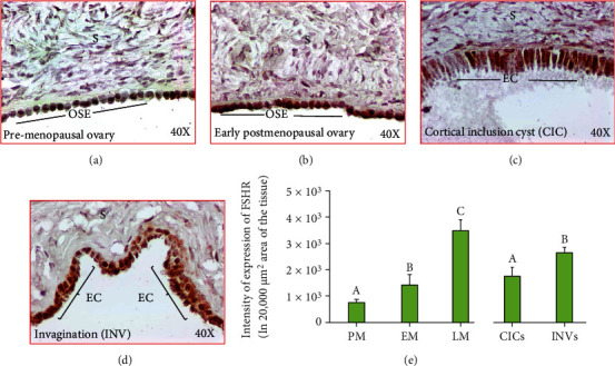
Expression of follicle-stimulating hormone receptor (FSHR) in ovaries during aging. (a)–(d): (a) Section of an ovary from a premenopausal subject showing a weak or moderate expression for FSHR by the ovarian surface epithelial (OSE) cells. (b) Section of an ovary from an early-stage postmenopausal subject showing strong expression for FSHR by the ovarian surface epithelial (OSE) cells. (c, d) Ovarian sections from healthy late postmenopausal women showing expression of FSHR by the epithelial cells (EC) in cortical inclusion cyst (CIC) and stromal invagination (INV), respectively. Compared with CIC, stromal INV showed stronger staining for FSHR expression. S: stroma; magnification: 40×. (e) Compared with premenopausal subjects, the intensities of FSHR expression were significantly (P < 0.04) higher in early menopausal women and increased further in late-stage menopausal women (P < 0.0001) (e). Furthermore, compared with CICs, the intensity of FSHR expression was significantly higher in INVs (P < 0.004) (e). y-axis shows mean ± SEM (n = 5 for each group) in 20,000 μm2 area of the tissue, and bars with different letters are significantly different. Details of statistical analysis are mentioned in materials and method section of the main text.