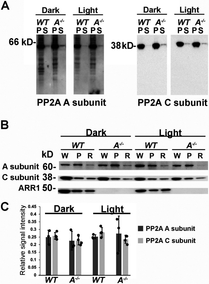
Figure 4.
PP2A levels and localization are not affected by ARR1 or light exposure. A, Western blot of membrane (P) and soluble (S) fractions of retinal homogenates from dark-adapted or light-exposed WT and Arr1−/− mice probed with antibodies against both isoforms of the A subunits or the C subunits. Equal amounts of proteins were loaded per lane. B, Western blots of whole retinal homogenate (W), the membrane fraction (P), or ROS (R) from dark-adapted or light-exposed WT or Arr1−/− mice probed with the indicated antibodies. C, Quantification of signals from PP2A A (N = 3) and C subunits (N = 4) in ROSs isolated from dark-adapted or light-exposed WT and Arr1−/− mice.

