
Figure 5.
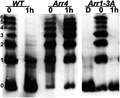
Rhodopsin dephosphorylation in transgenic mice expressing different visual arrestins in rods. Representative image from three independent experiments. C57 WT and mice that express the ARR4 or ARR1-3A transgenes on the Arr1−/− background were exposed to light and returned to darkness for 1 h. The status of rhodopsin phosphorylation was examined by IEF. Left, The number of rhodopsin-attached phosphates.