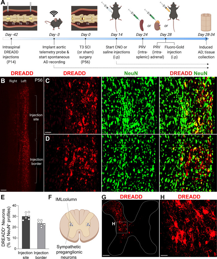
Figure 1.
Intraspinal injections of Gi DREADDs transduce VGluT2+ spinal interneurons. A, Experimental timeline. Mice received intraspinal DREADD injections at T4 and T7 at P14. Six weeks later, mice were implanted with radiotelemetry probes, followed by T3 laminectomy (sham) or T3 SCI. Mice received 1–3×/d CNO or saline injections from day 14 to the end of the study. Mouse cohorts were injected with intrasplenic PRV at day 24, intra-adrenal PRV at day 28, or intraperitoneal Fluoro-Gold at day 28, then were perfused at day 28, 32, and 34, respectively (see figure legends for individual experiment details). B, Representative image of T4–T6 horizontal spinal cord section at the level of the central canal (IML level), showing VGluT2+ interneurons that have been induced by Gi DREADD (red profiles). Scale bar, 200 μm. C, D, High-magnification representative images of an injection site (C) and an injection border site (D), showing the density of DREADD-induced VGluT2+ (red) interneurons relative to total neurons (NeuN+ profiles). Scale bar, 100 μm. E, Quantification of DREADD-induced neurons as a percentage of total NeuN+ neurons in the intermediate gray matter of the thoracic spinal cord. Transduction efficiency decreases slightly away from the injection site. n = 6 mice/group; values are the mean ± SEM. F–H, Diagram showing IML neuron location and representative images of T7 spinal cord coronal section showing dorsal–ventral transduction of Gi DREADD. Transduced neurons are primarily in spinal laminae III–VIII and X. Scale bars: G, 200 μm; H, 40 μm.

