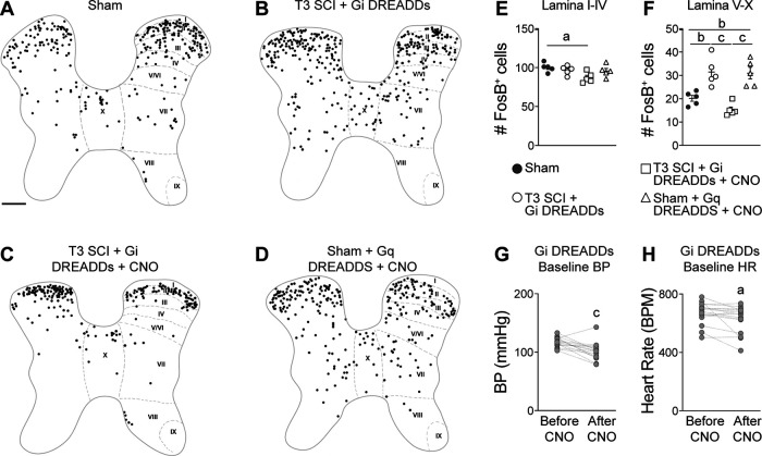
Figure 3.
Silencing VGluT2+ interneurons reduces neuron activity in the thoracic spinal cord. A–D, Representative binarized spinal cord cross sections (spinal level T7) immunostained for FosB, a marker of activated neurons. FosB+ cells were marked with circles of uniform size for clarity. Scale bar: A–D, 200 μm. E, F, Quantification of FosB+ cells in laminae I–IV (E) and laminae V–X (F). One-way ANOVA with Tukey's post hoc tests; n = 4–5 mice/group. ap < 0.05; bp < 0.01; cp < 0.001; values are the mean ± SEM. G, H, The resting blood pressure (G) and heart rate (H) of VGluT2-cre mice injected with Gi DREADDs before and after CNO injection. Paired Student's two-sided t tests, n = 16 mice/group. ap < 0.05; cp < 0.001 versus before CNO injection. Anatomical mapping of FosB+ and DREADD+ neurons is shown in Extended Data Figure 3-1.

