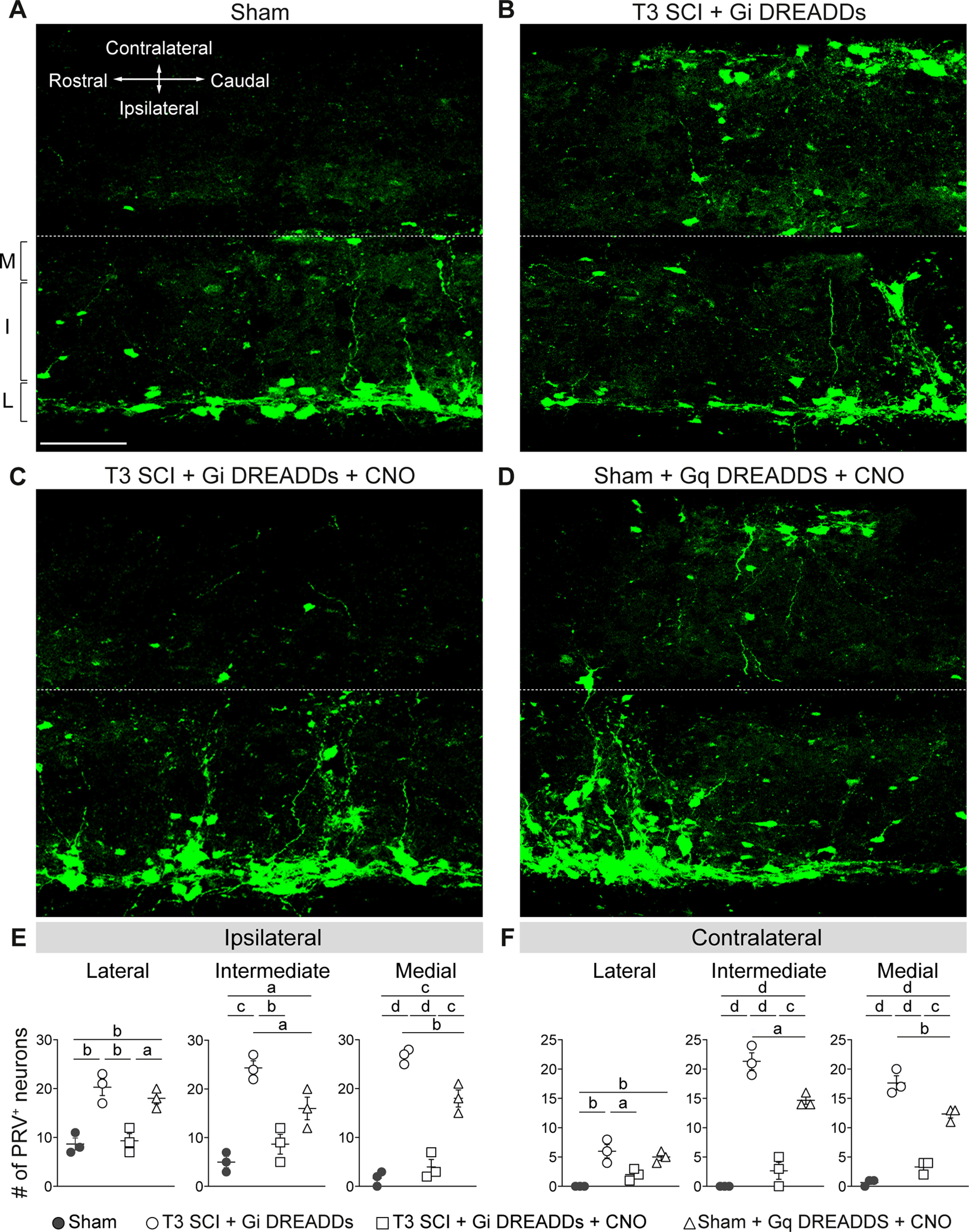
Figure 7.

Silencing VGluT2 interneurons prevents expansion of the sympathetic spinal–splenic circuitry. At 24 dpi, PRV-GFP was injected into the spleen for retrograde labeling of the spinal–splenic sympathetic innervation. At 4 d postinjection (28 dpi), animals were perfused and spinal cords analyzed for PRV+ neurons. M, Medial; I, intermediate; L, lateral gray matter. A–D, Representative horizontal sections of the thoracic spinal cord (T7). Scale bar, 100 μm. E, F, Quantification of PRV+ neurons in the ipsilateral (E) and contralateral (F) lateral, intermediate, and medial zones of T7 gray matter. One-way ANOVA with Tukey's post hoc tests; n = 3 mice/group. ap < 0.05; bp < 0.01; cp < 0.001; dp < 0.0001. Values are the mean ± SEM.
