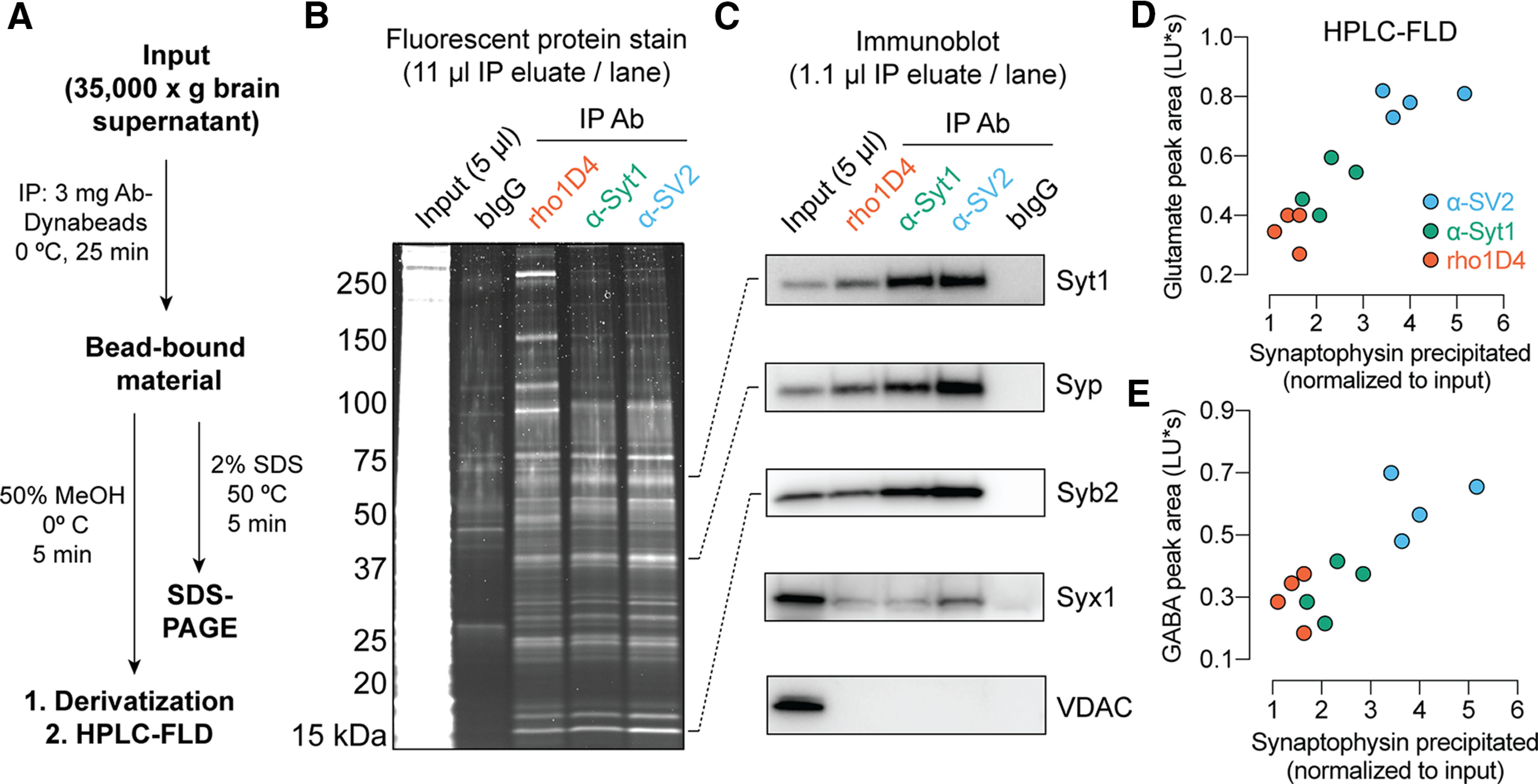
Figure 1.

Immunopurification of synaptic vesicles. A, Scheme for vesicle immunoprecipitation and analysis. Each mouse brain provided sufficient material for analysis of protein and neurotransmitter using two different mAbs. B, Staining of proteins separated by SDS-PAGE demonstrates broad similarity among anti-SV2, anti-syt1, and rho1D4 immunoprecipitates, with minimal protein binding by control beads bearing pooled bovine IgG. Note the dominant band at 38 kDa, corresponding to synaptophysin. C, Immunoblot analysis of precipitated material. Each antibody yields strong enrichment of SV proteins, but only the expected weak enrichment of the plasma membrane t-SNARE syntaxin-1 and no detectable contamination from the mitochondrial protein VDAC. D, E, For each experiment, the area of the HPLC fluorescence peak corresponding to NBD-derivatized glutamate (D) or GABA (E) was plotted against the normalized intensity of the synaptophysin band on immunoblot (n = 4 biological replicates using two separately prepared batches of Ab-Dynabeads for each mAb; B–E). Example raw chromatograms used for GABA and glutamate measurements are shown in Extended Data Figure 1-1. “LU*s”, luminance units * seconds.
