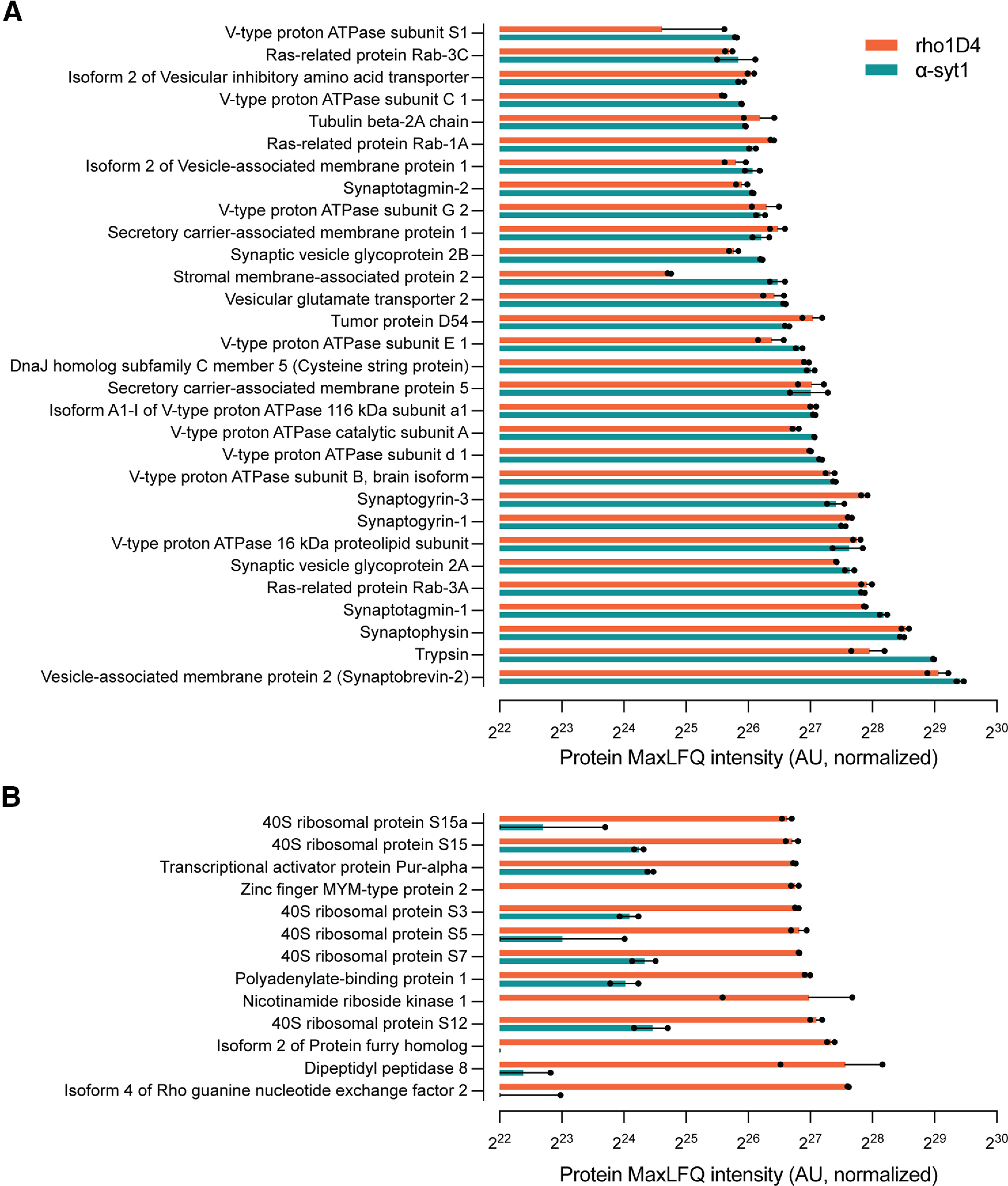
Figure 2.

Proteomic characterization of α-syt1 and rho1D4 immunoprecipitated SVs. A, Proteins identified by Q-TOF LC-MS after bead elution with SDS were sorted by intensity in the α-syt1 immunoprecipitates (n = 2 biological replicates per immunoprecipitation antibody). Intensity values were calculated using the IonQuant function and MaxLFQ algorithm in the FragPipe analysis software. The proteins with the top 30 highest intensity scores in α-syt1 IP samples, not including antibody fragments, are shown here. The majority of identified proteins are well known to reside on SVs and are well matched in intensity by the rho1D4-precipitated material, confirming that the rho1D4 mAb effectively immunoprecipitates a population of SVs similar to those obtained with the α-syt1 antibody. B, Proteins among the top 30 in SDS-eluted rho1D4 IP samples that were not observed among the top 30 in α-syt1 IP samples. Source data used to generate this figure are shown in Extended Data Figure 2-1.
