Figure 3.

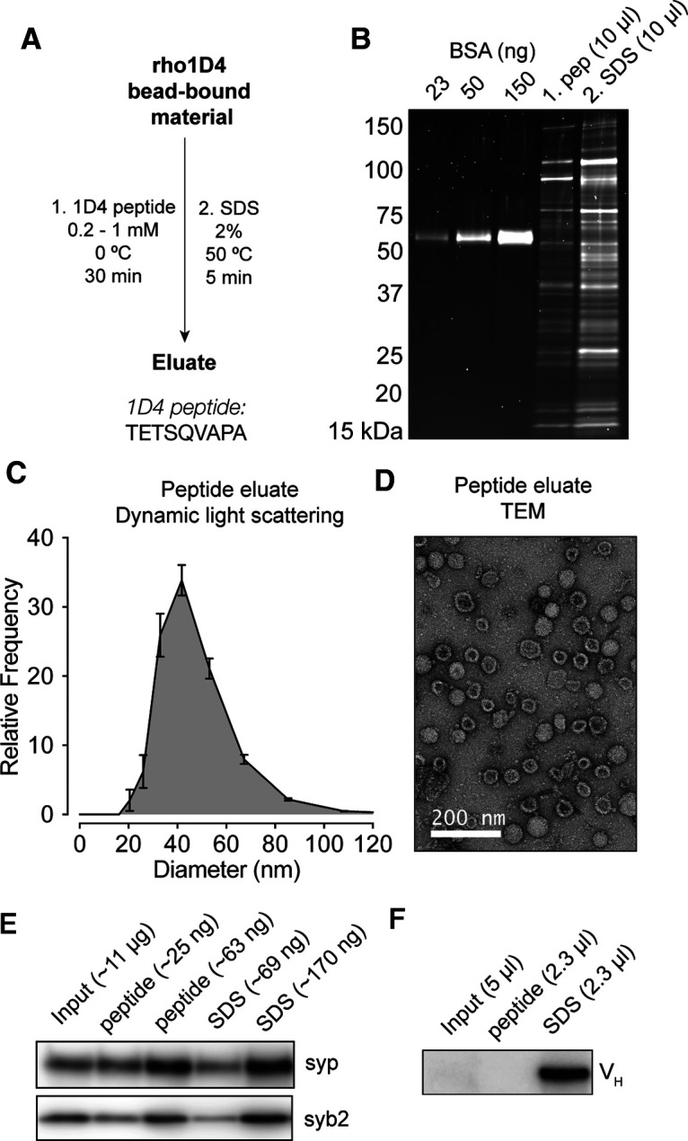
rho1D4-IP enables peptide elution of native SVs. A, Purification scheme for peptide elution. The amino acid sequence of the 1D4 peptide sequence is shown. B, SDS-PAGE with fluorescent stain of protein eluted from rho1D4 beads using the 1D4 peptide followed by 2% SDS. SV proteins were readily eluted from the beads with the 1D4 peptide. C, Dynamic light scattering measurements (n = 3 biological replicates) of eluted material indicates a single population of particles ∼40–50 nm in diameter. This population represented >99% of particles detected in each sample. D, Negative-stain TEM of 1D4-eluted material demonstrates vesicles of the appropriate size, decorated with expected structures, with minimal contamination by nonvesicular structures. E, Immunoblot of SV proteins in input fraction along with peptide and SDS eluates from rho1D4 beads, with approximate amount of total protein loaded per lane. F, Immunoblot of peptide and SDS eluates probed with anti-mouse secondary mAb demonstrating the relative absence of eluted mAb in peptide eluates.
