. 2020 May 19;10(32):18959–18969. doi: 10.1039/c9ra03736e

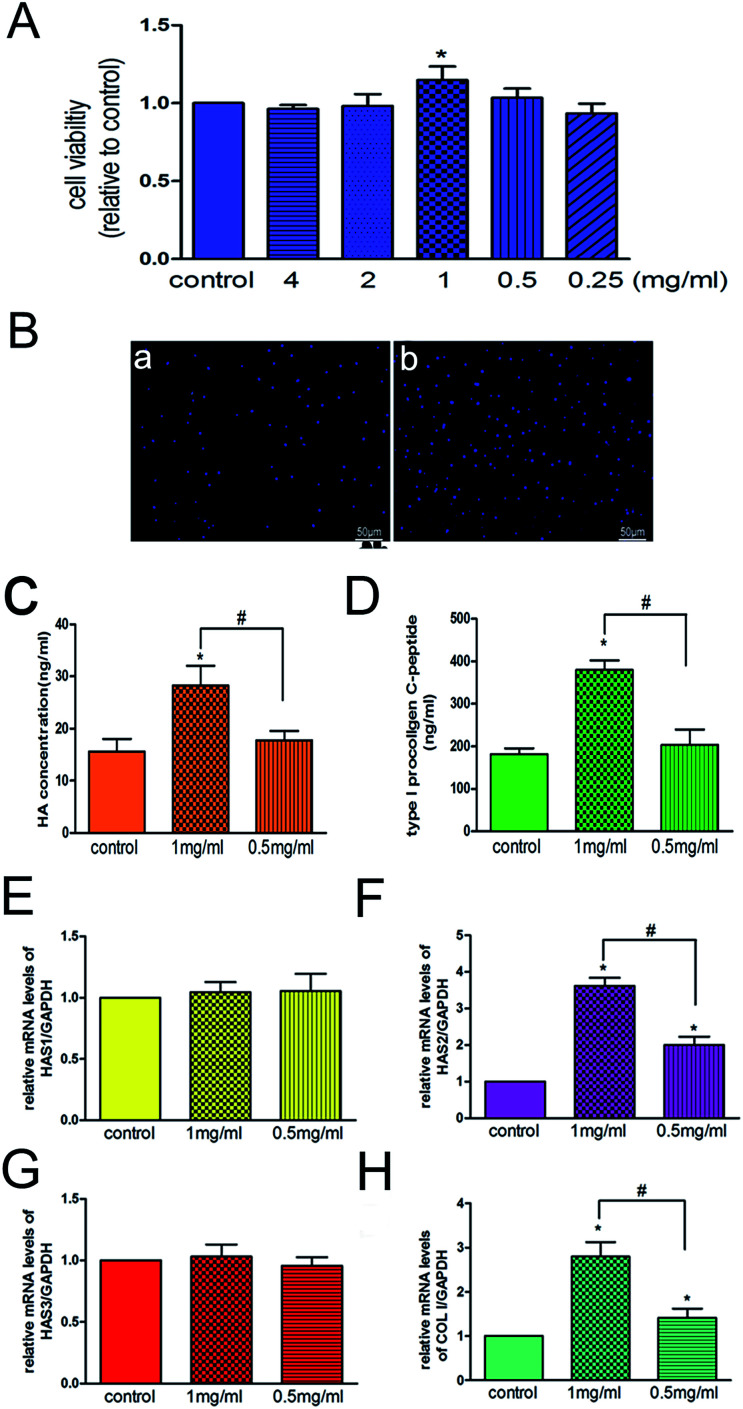
Fig. 5. (A) HSFs were treated with various concentrations of 3D-GF-PADM. MTT results showed that 1 mg ml−1 3D-GF-PADM enhanced the proliferation of HSFs. (B) 4,6-Diamidino-2-phenylindole (DAPI) staining was used to analyse the effect of 3D-GF-PADM treatment on cell viability. A significant difference was observed between the 3D-GF-PADM treatment group and the control group. (C) The increased HA secretion induced by 1 mg ml−1 3D-GF-PADM. (D) The increased collagen secretion induced by 1 mg ml−1 3D-GF-PADM. (E) The relative HAS1 mRNA expression after treatment with 3D-GF-PADM. There was no significant change compared with the control group. (F) The relative HAS2 mRNA expression after treatment with 3D-GF-PADM. After treatment with 1 mg ml−1 3D-GF-PADM, the expression of HAS2 was increased. (G) The relative increase of HAS3 mRNA expression after treatment with 3D-GF-PADM. (H) The relative COL I mRNA expression after treatment with 3D-GF-PADM.

Fig. 5