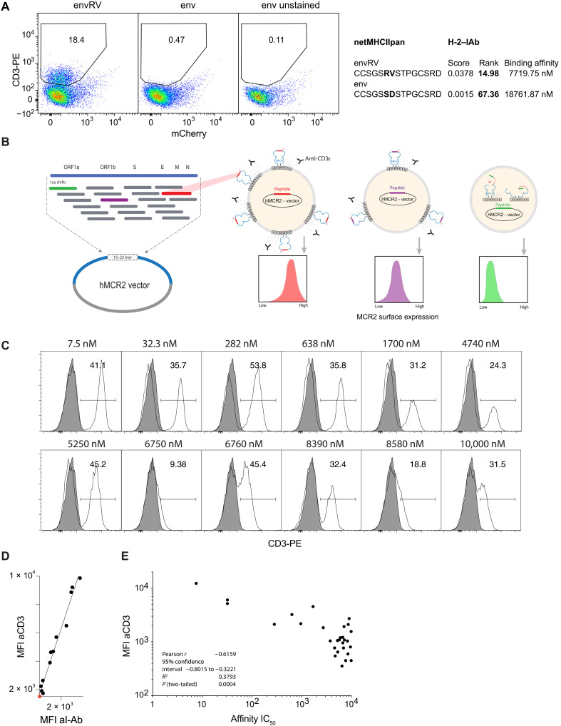
Fig. 1. Cell surface expression of MCR2 depends on peptide-MHC binding.
(A and C) Flow cytometric analysis of CD3ε expression on 16.2c11 cells transduced with (A) MCR2-envRV or MCR2-env or (C) MCR2 constructs carrying peptides binding I-Ab with different affinity (7.5 nM to 10 μM). In (A), the x axis shows irrelevant, background nuclear factor of activated T cell (NFAT)–mCherry reporter reactivity. Gray histograms, untransduced cells. PE, phycoerythrin. (B) Schematic representation of the peptide library cloning into the human MCR2 vector and of the MEDi principle. ORF1a, open reading frame 1a; aa, amino acid. (D) Comparison of MFI of anti-CD3ε and anti–I-Ab stainings. (E) Correlation of MFI of anti-CD3ε staining (of MCR2+ cells) with the peptide-MHC binding affinity of peptides carried by the MCR2.

