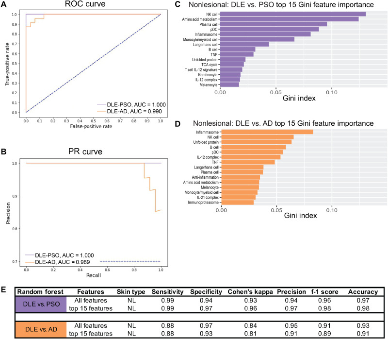
Fig. 6. Nonlesional DLE is distinct from PSO and AD.
(A) ROC curve and (B) PR curve of nonlesional DLE samples compared to nonlesional PSO (purple) samples and nonlesional DLE samples compared to nonlesional AD samples (orange) using all cellular and pathway gene signatures. Top 15 features important in classifying (C) nonlesional DLE and nonlesional PSO and (D) nonlesional DLE and nonlesional AD using Gini feature importance. (E) Classification metrics to properly separate nonlesional DLE samples and nonlesional PSO or nonlesional AD samples using all 48 (top) or the top 15 (bottom) cellular and pathway gene signatures. Refer to table S3 (A and B) for ML details. Collinear features were removed (fig. S15).

