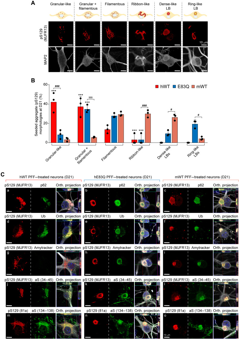
Fig. 8. Comparative analysis of the morphological properties and diversity of aSyn aggregates and inclusions formed at 21 days after treatment of hippocampal neurons with human E83Q and WT PFFs and mouse WT PFFs.
(A) LB-like pathological diversity was observed in PFF-treated hippocampal neurons. The representative images for the filamentous and the ribbon-like inclusions shown in this panel are also depicted in figs. S13 to S16 with additional markers of the LB-like inclusions. (B) Quantification of the morphologies of the aSyn seeded aggregates observed by ICC in primary hippocampal neurons at D21. A minimum of 100 neurons was counted in each experiment (n = 3). The graphs represent means ± SD of a minimum of three independent experiments. ***P < 0.0005 (ANOVA followed by Tukey post hoc test, mWT PFF–treated neurons versus human PFF (hWT or E83Q)–treated neurons). ###P < 0.0005 (ANOVA followed by Tukey post hoc test, hE83Q PFF–treated neurons versus mWT PFF– or hWT PFF–treated neurons). $$$P < 0.0005 (ANOVA followed by Tukey post hoc test, hWT PFF–treated neurons versus hE83Q PFF–treated neurons). #P < 0.05. (C) Aggregates were detected by ICC using pS129 (MJFR13 or 81a) in combination with p62 (a to c), ubiquitin (Ub) (d to f), Amytracker dye (g to i), total N-terminal aSyn (epitope, 34 to 45) (j to l), or total C-terminal (epitope, 134 to 138) (m to o) aSyn antibodies in hWT PFFs (a, d, g, j, and m), hE83Q PFFs (b, e, h, k, and n), or mWT PFF–treated hippocampal neurons (c, f, i, l, and o). Neurons were counterstained with MAP2 antibody, and the nuclei were counterstained with DAPI. Scale bars, 10 μm.

