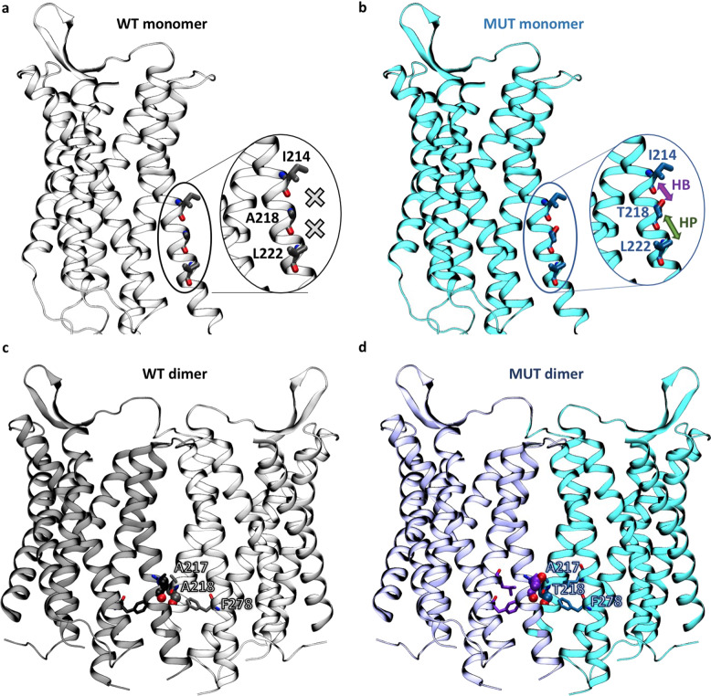
Fig. 3. Comparison of the local environment of residue 218 in wild-type (WT) and mutant (MUT) models based on the crystal structure of the OXTR A218T variant (PDB code 6TPK).
a, b Monomeric forms (inactive state). No interactions, hydrogen bonds (HBs) and hydrophobic interactions (HPs) involving the residue are indicated by a cross, a violet arrow, and a green arrow, respectively. The counterpart homology models of the active and intermediate states are shown in the Supplementary Information (Fig. S2). c, d Top homodimeric models with a TM5 interface based on the experimental structure of the μ-opioid receptor dimer (PDB code 4DKL). The residue in position 218 is shown as spheres and residues surrounding it within 5.5 Å as sticks. The other OXTR/OXTR models are shown in the SI (Fig. S3).

