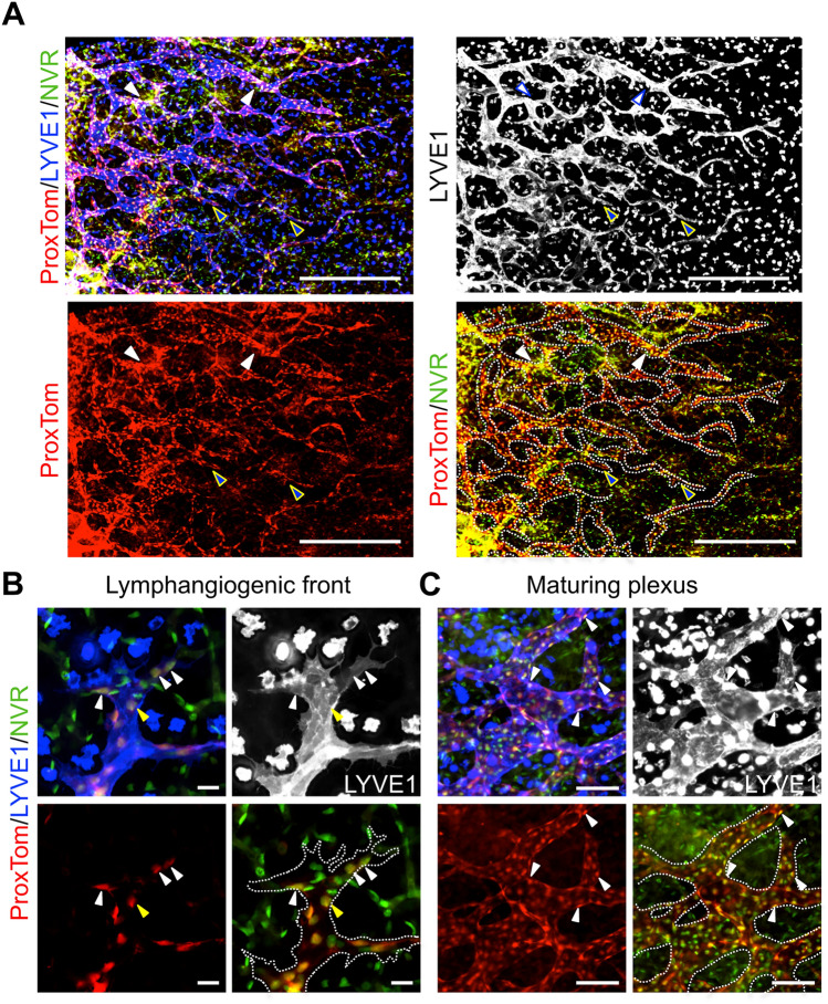
Fig. 2.
Notch activation observed throughout the embryonic dermal lymphatic vascular plexus. E14.5 ProxTom;NVR skin wholemounts stained for LYVE1. a Low magnification image demonstrating Notch activity throughout the developing lymphatic plexus. Blue arrowheads mark sprouts at the lymphangiogenic front with Notch activity. White arrowheads mark regions of high Notch signaling in the maturing plexus. Scale bars, 500 μm. b High magnification of spiky-ended lymphatic sprout. White arrowheads mark tip cells with Notch activity. Yellow arrowheads mark stalk cells with Notch activity. c High magnification of the maturing plexus. White arrowheads mark LECs with Notch activity. b, c Scale bars, 100 μm

