Fig. 6.
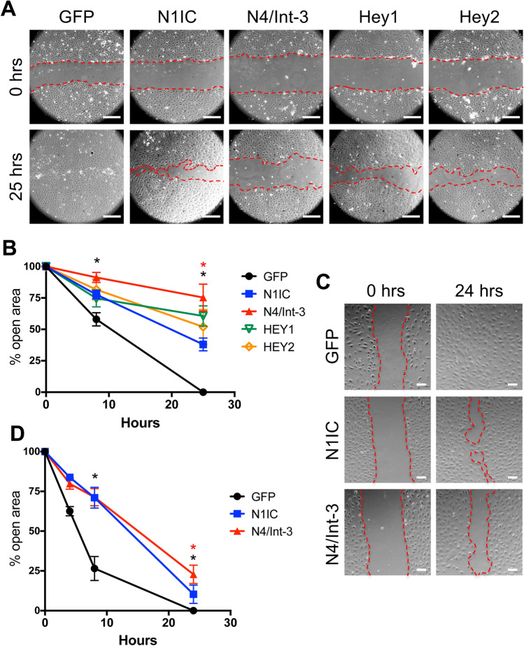
Notch signaling inhibited LEC migration. a Confluent N1IC-, N4/Int-3-, Hey1-, Hey2-, or GFP-expressing HdLECs were scratched and representative images for 0 and 25 h presented. Scale bars, 2.5 μm. b Quantification of percent open wound area at 0, 8, and 25 h. Data presented ± s.e.m. two-way-ANOVA: p < 0.0012, t test: *p < 0.002 N1IC or N4/Int-3 vs. GFP at 8 and 25 h, *p < 0.002 N4/Int-3 vs. N1IC at 25 h. c Confluent N1IC-, N4/Int-3-, or GFP-expressing HdLECs were treated with mitomycin C and scratched and representative images for 0 and 24 h presented. Scale bars, 100 μm. d Quantification of percent open wound area at 0, 4, 8, and 24 h. Data presented ± s.e.m. two-way ANOVA: p < 0.0001, Dunnett’s multiple comparison test *p < 0.0001 N1IC or N4/Int-3 vs. GFP. t test: *p < 0.0001 N4/Int-3 vs. N1IC

