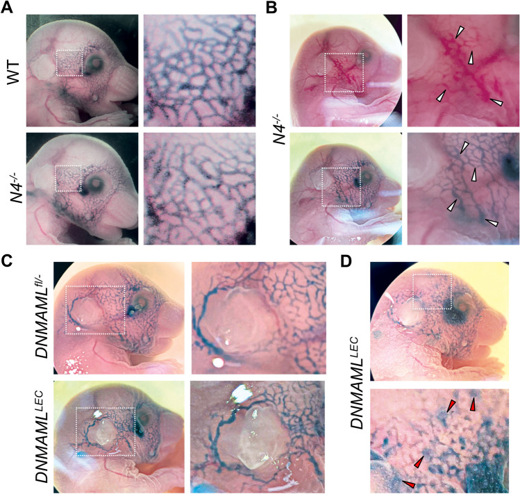
Fig. 8.
Loss of Notch4 and deletion of LEC canonical Notch signaling resulted in distinct lymphatic phenotypes at E17.5. a, b Lymphangiography of E17.5 wild-type (n = 6) and Notch4−/− (n = 8) embryos. Representative images of wild-type and Notch4−/− embryos. Boxed area enlarged to the right. b Notch4−/− embryos with blood-filled dermal lymphatics (white arrowheads). Boxed area enlarged to the right. c, d Prox1CreERT2 and DNMAMLfl/fl mice were crossed and tamoxifen administered at E12.5 and lymphangiography performed at E17.5. Control (n = 12), DNMAMLLEC (n = 5). c Representative images of DNMAMLfl/− control and DNMAMLLEC embryos. Boxed area enlarged to the right. d DNMAMLLEC embryo with leaking dermal lymphatic vessels (red arrowheads). Boxed area enlarged below

