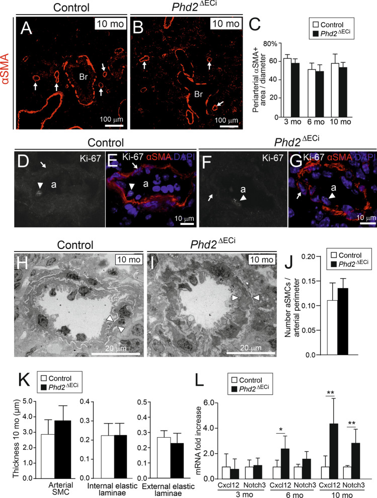
Fig. 3.
Lack of arterial smooth muscle proliferation, arterial wall and perivascular matrix remodeling in Phd2ΔECi pulmonary arteries/arterioles. A and B Lung sections of 10-month-old mice immunostained with alpha smooth muscle actin (αSMA). Arrows point to αSMA positive arterioles; bronchus (Br). C Area of αSMA positive cells around arteries and arterioles, n = 5 mice/group, 10 arteries/mouse calculated. D–G, Arterioles (a) stained with Ki-67, αSMA and DAPI. EC and aSMCs nuclei are indicated by arrowheads and arrows, respectively. There was no change in proliferation marker Ki-67 between the genotypes. H and I Representative TEM micrographs of pulmonary arterioles, thickness of single aSMC is indicated by arrowheads. J Number of aSMCs/perimeter of arteriole. K Thickness of arterial layers in 10-month-old mice. n = 5–7 mouse/group (1–4 arteries/mouse). L Expression of SMCs signaling molecules Cxcl12 and Notch3 in the lungs. n = 5–8 mice/group. Mean ± SD. *P < 0.05, **P < 0.01 in t-test. Mo, month

