. 2020 Jul 15;10(45):26594–26603. doi: 10.1039/d0ra04648e

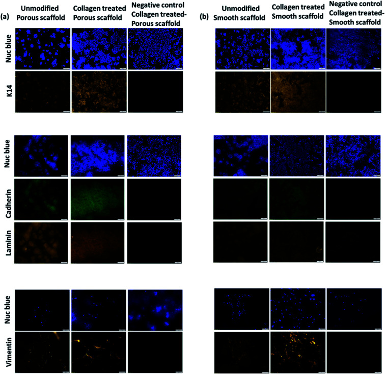
Fig. 6. Immunofluorescence of co-cultured HaCaT and HFF cells in electrospun PLLA scaffolds. Panel (a) corresponds to cells cultured in porous scaffold including unmodified scaffold and 0.1% collagen modified scaffold. A negative control was maintained without the treatment of primary antibodies. HaCaT cells were stained with primary antibodies for cytokeratin 14, cadherin, laminin V and HFF cells were stained with primary antibodies for vimentin, followed by the treatment of fluorescently labelled secondary antibodies. Corresponding nuclei were stained with NucBlue. Panel (b) corresponds the same immunofluorescence staining for HaCaTs and HFF cells in unmodified smooth and collagen modified smooth PLLA scaffolds (scale bar = 100 μm).

Fig. 6