. 2020 Jul 21;10(45):27064–27080. doi: 10.1039/d0ra03194a

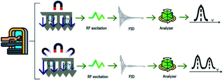
Fig. 3. To obtain an MRI image, the body is located in a uniform magnetic field. Hydrogen nuclei align with the magnetic field and create a net magnetic moment parallel to it. When a radio-frequency pulse (RF excitation) is applied perpendicularly, the net magnetic moment of the nuclei tilts away from the magnetic field. When the RF pulse stops, the nuclei return to the equilibrium initial state (relaxation step). During the relaxation, the nuclei lose energy and a measurable signal, indicated as the free-induction decay (FID), is detected. 3D MRI images can be generated, encoding the FID in each dimension. An additional magnetic field in the gradient changes the FID as a function of the proton 3D position.

Fig. 3