. 2020 Jul 21;10(45):27064–27080. doi: 10.1039/d0ra03194a

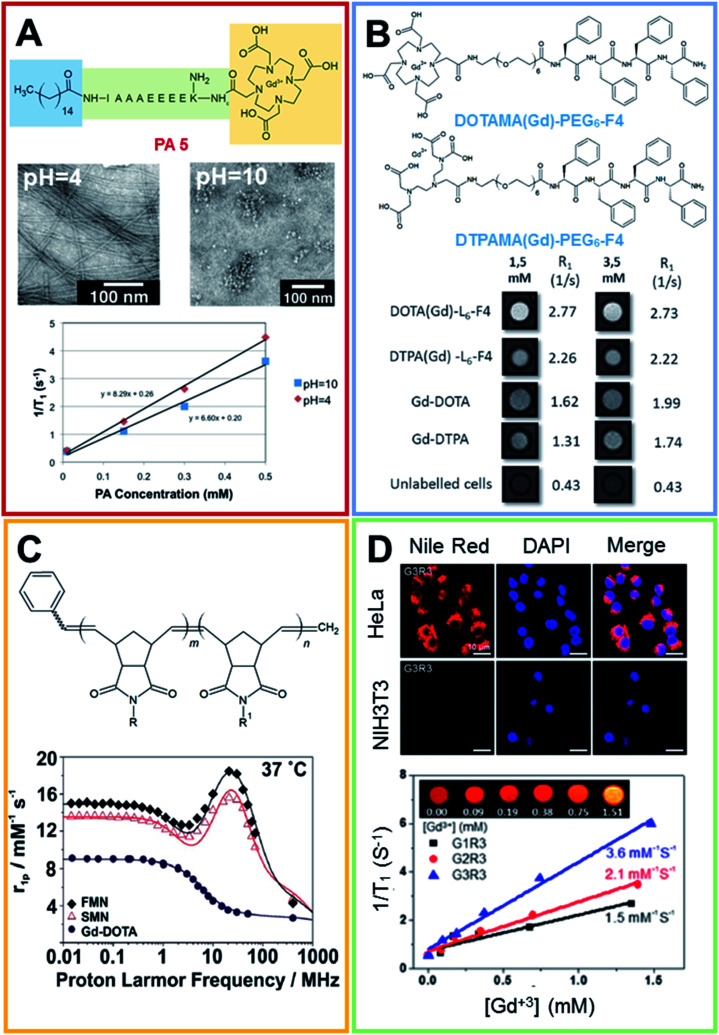
Fig. 4. (A) Chemical formula for the PA 5 compound, the peptide sequence of PA 5 is reported according to the one code letter. TEM images (0.5 mM solution of PA 5) at pH 4.0 (on the left) and 10.0 (on the right). T1 relaxivity graph of PA 5 under basic and acidic conditions (adapted with permission40 2012-American Chemical Society). (B) Chemical structure of DOTAMA(Gd)-PEG6-F4 and DTPAMA(Gd)-PEG6-F4. T1-weighted MR-images of pellets of the J774A.1 cell line labelled with 1.5 and 3.5 × 10⁻³ mol L−1 of the two Gd-complexes. The relative observed relaxation rates are reported too (reproduced with permission from ref. 41). (C) Chemical structure of Gd3+-containing an amphiphilic block copolymer. R and R′ can be a phenyl group or a modified Gd-DOTA complex (norbornenyl-Gd-DOTA monoamide). 1H NMRD profiles for Gd-DOTA, spherical and fibrillar micellar nanoparticles (reproduced with permission,51 published by The Royal Society of Chemistry). (D) Laser scanning microscopy images of HeLa and NIH3T3 cells lines treated with Nile red-loaded porous networks of Gd3+-G3R3 (1 h after incubation): left, Nile red (red); middle, nuclei stained with DAPI (blue); and right, merged images. T1 relaxivity plots of Gd3+-chelating nanostructures (0.2 mM) as a function of the Gd3+ concentration ([Gd3+]) (4.7 T, 25 °C) and (inset) T1-weighted MR images of Gd3+-G3R3 (adapted with permission,50 copyright 2017-American Chemical Society).

Fig. 4