. 2020 Jul 21;10(45):27064–27080. doi: 10.1039/d0ra03194a

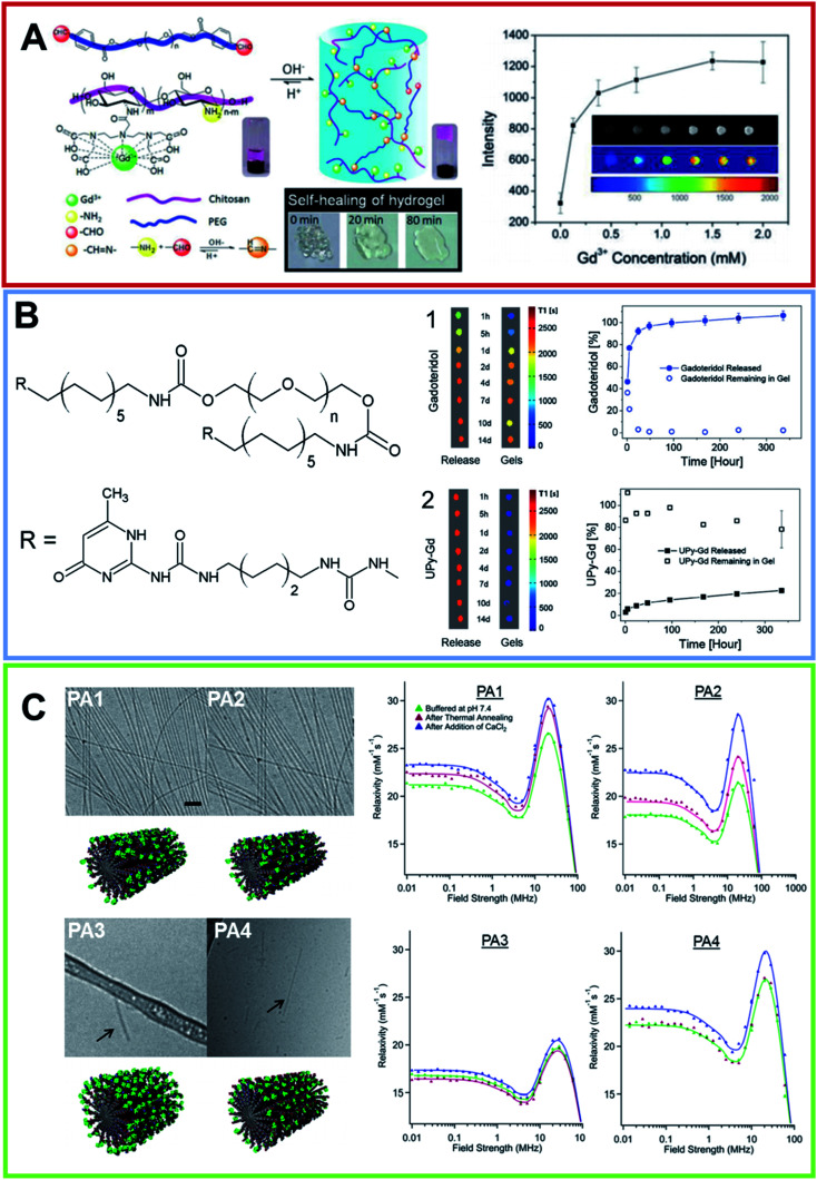
Fig. 5. (A) On the left, schematic illustration of the formation of pH-sensitive injectable hydrogel composed by a modified PEG polymer (blue ribbon) and a DTPA-decorated chitosan (purple ribbon). The hydrogel self-healing feature is reported too. On the right, MRI intensity of the visible hydrogel as a function of the Gd(iii) concentration (from ref. 57. Reproduced by permission of The Royal Society of Chemistry). (B) Chemical structure for the bifunctional hydrogelator containing ureidopyrimidinone (UPy). 10 wt% UP-PEG hydrogels T1-weighted 1.5 T MRI scans reported in pseudo-color of the released solution and for the gels. Hydrogels contain either (1) 1 × 10⁻³ mol L−1 Gadoteridol or (2) 1 × 10⁻³ mol L−1 UPy-Gd(iii) modified monomers. The relative release curves are reported too (arranged with permission59). (C) On the left, Cryo-TEM of PAs conjugates in 10 mmol L−1 in Tris buffer after a process of thermal annealing (80 °C for 30 min) and their relative molecular graphical representation. The scale bar represents 200 nm and Gd macrocycles are represented as green elements. On the left, the NMRD profiles for all PAs at three different conditions recorded at 37 °C and a PAs concentration of 2 mmol L−1 (adapted with permission,67 copyright 2014-American Chemical Society).

Fig. 5