
Figure 5.
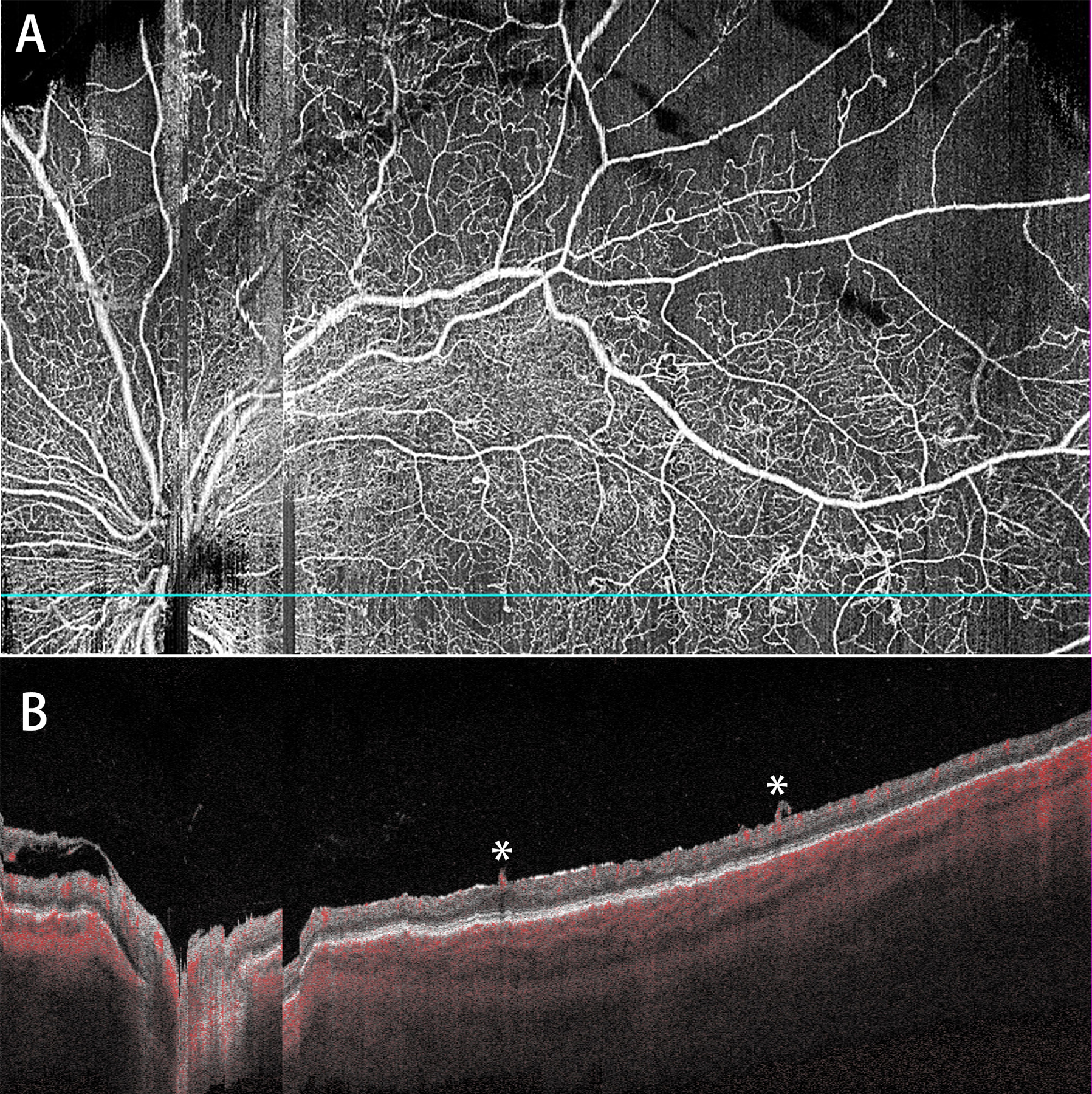
Representative WF SS-OCTA images of a PDR eye with the occurrence of VH during the follow-up (28 days) (A: whole retina slab; B: corresponding B-scan). Two forward NVs (*) are shown in B.
Official websites use .gov
A
.gov website belongs to an official
government organization in the United States.
Secure .gov websites use HTTPS
A lock (
) or https:// means you've safely
connected to the .gov website. Share sensitive
information only on official, secure websites.
Representative WF SS-OCTA images of a PDR eye with the occurrence of VH during the follow-up (28 days) (A: whole retina slab; B: corresponding B-scan). Two forward NVs (*) are shown in B.