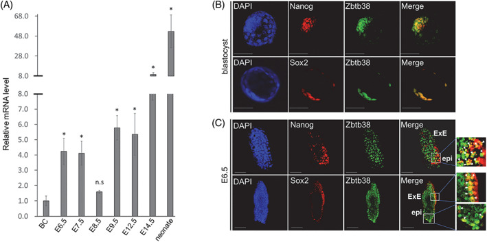
FIGURE 1.

Expression of Zbtb38 during mouse embryonic development. (A) Results of the fold change of qRT–PCR for Zbtb38 expression during mouse embryonic development. The graph shows the fold change relative to blastocyst, which is denoted by 1. Embryos from the indicated stages were carefully isolated from the uterus of C57BL/6J mice intercrosses. The TATA‐binding protein (TBP) gene was used as an internal control, and the levels of transcripts were normalized against the TBP gene. Data are representative of three independent replicates measured in triplicates, and error bars indicate ± S.D. *p < 0.05. ‘n.s.’ indicates no significance. (B and C) Whole‐mount immunofluorescence and confocal microscopy for blastocyst (B) and E6.5 (C). Expressions of the indicated proteins were detected using anti‐ Zbtb38, anti‐Nanog and anti‐Sox2 antibodies. Cell nuclei were counterstained with DAPI. White arrows indicate regions of colocalization. Scale bar denotes 50 μm (1B) or 100 μm (1C). epi, epiblast; ExE, extraembryonic ectoderm
