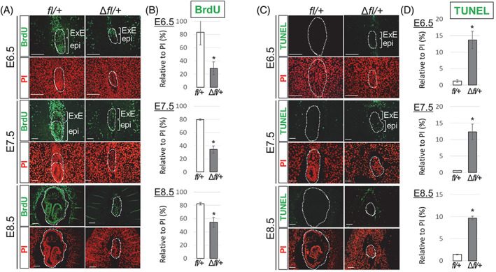
FIGURE 4.

Evaluation of proliferating and apoptotic cells in E6.5–E8.5 embryos. (A and B) Heterozygous loss of Zbtb38 inhibits epiblast cell proliferation of E6.5–E8.5 embryos. (A) Immunofluorescence analysis of paraffin‐embedded sections of controls and Zbtb38 ∆fl/+ embryos at E6.5–E8.5. Two consecutive paraffin‐embedded sections were taken for performing immunostaining with anti‐BrdU antibody (green), and nuclei were counterstained with PI (red). epi, epiblast; ExE, extraembryonic ectoderm. Scale Bar: 50 μm. (B) Quantitative analysis of the number of labelled BrdU cells relative to the total number of PI‐positive nuclei from the indicated umbers of embryos at E6.5 (fl/+: n = 9; ∆fl/+: n = 8), E7.5 (fl/+: n = 11; ∆fl/+: n = 9), and E8.5 (fl/+: n = 6; ∆fl/+: n = 5). Error bars represent ± S.E.M. *p < 0.05. (C and D) Heterozygous loss of Zbtb38 induces apoptosis of E6.5–E8.5 embryos. (C) A TUNEL assay was performed on paraffin‐embedded sagittal sections from E6.5 embryos onwards. TUNEL‐positive cells are shown in green, and nuclei were counterstained with PI (red). Scale Bar: 50 μm. (D) Quantitative analysis of the number of TUNEL‐positive cells relative to the total number of PI‐positive nuclei from the indicated umbers of embryos at E6.5 (fl/+: n = 9; ∆fl/+: n = 7), E7.5 (fl/+: n = 11; ∆fl/+: n = 8), and E8.5 (fl/+: n = 7; ∆fl/+: n = 5). Error bars indicate ± S.E.M. *p < 0.05
