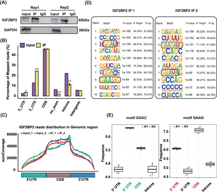
FIGURE 3.

RNA immunoprecipitation sequencing analysis of insulin‐like growth factor 2 mRNA‐binding protein 2's (IGF2BP2) binding profile and binding motifs. (A) IGF2BP2 protein detection by western blot in KGN cells. (B) Read distribution across the reference genome. Error bars represent the mean ± SEM. *P < 0.05, **P < 0.01, ***P < 0.001. (C) The peak reads density for 5′UTR, CDS and 3′UTR for input and IGF2BP2 IP samples. (D) Motif analysis using the software HOMER showing the top 10 preferred binding motifs of IGF2BP2. (E) The binding sites of the GGAC and GAAG binding motifs
