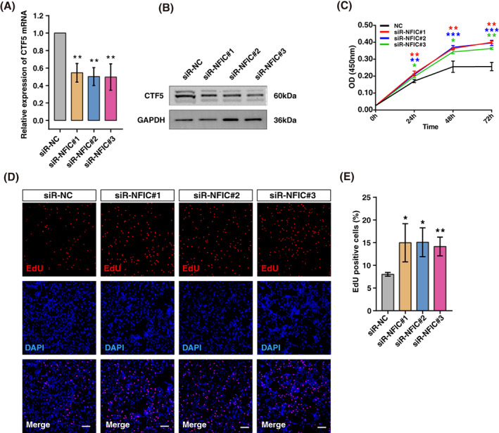
FIGURE 7.

Knockdown of nuclear factor 1 C‐type (NFIC) promotes cell proliferation in KGN cells. (A) The relative expression of CTF5 mRNA is detected following NFIC knockdown in KGN cells. **P < 0.01. (B) Western blot analysis of CTF5 following NFIC knockdown. (C) KGN cells were transfected with siRNA‐NC or siRNA‐NFIC#1/ #2/ #3 for 24, 48, or 72 h, and cell viability was determined by the CCK‐8 assay. *P < 0.05, **P < 0.01, ***P < 0.001. (D) EdU staining of NFIC knockdown cells. Nuclei were stained by using DAPI. EdU positive cells, red; cell nuclei, blue; the data shown were representative of three independent experiments with similar results. Scale bar, 100 μm. (E) Statistics of EdU positive cells quantified by counting the cells with fluorescent signal using the software Image J. Experiments were performed in triplicate. *P < 0.05, **P < 0.01
