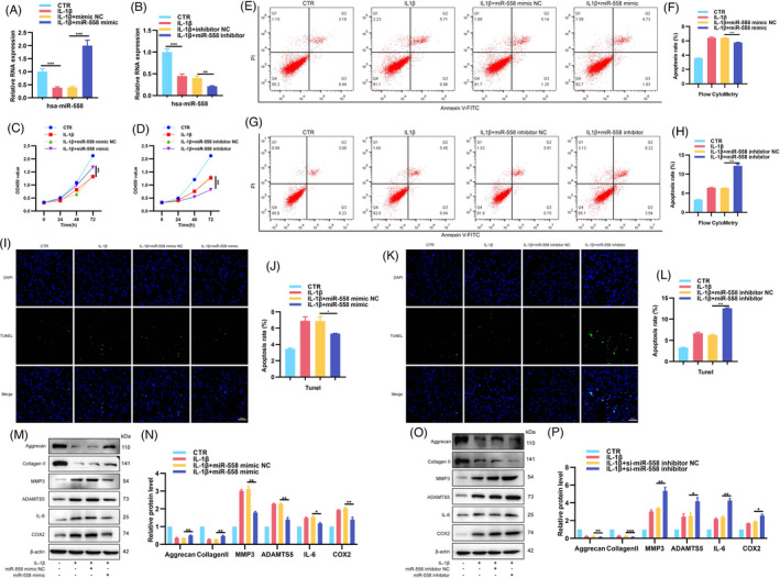
FIGURE 4.

Roles of miR‐558 in the proliferation, apoptosis, ECM synthesis and degradation, and inflammatory factor release in NP cells. (A, B) NP cells treated with IL‐1β were transfected with miR‐558 mimic, inhibitor or NC, and levels of miR‐558 were detected by RT‐qPCR. (C, D) CCK8 assay examined proliferation of NP cells treated by IL‐1β with or without miR‐558 mimic or miR‐558 inhibitor. (E–L) Flow cytometry and TUNEL assay were used to detect NP cells apoptosis. (M–P) NP cells were transfected with miR‐558 mimic, inhibitor or NC before IL‐1β stimulation, and protein levels of aggrecan, collagen Ⅱ, MMP3, ADAMTS5, IL‐6 and COX2 were detected. *p < 0.05, **p < 0.01 and ***p < 0.001 vs. the indicated group. Statistical data were presented as mean ±SEM; FITC, fluorescein isothiocyanate; PI, propidium iodide; TUNEL, terminal dexynucleotidyl transferase (TdT)‐mediated dUTP nick‐end labelling; DAPI, 4′,6‐diamidino‐2‐phenylindole
