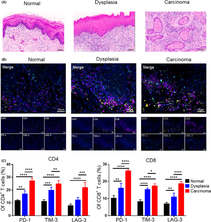
FIGURE 6.

Expression of inhibitory receptors on T cells increased during the development of human OSCC. (A) Representative hematoxylin and eosin (H&E) staining images of human normal, dysplasia, and carcinoma tissues. (B) Representative multiplex immunohistochemistry (mIHC) images showing expression of inhibitory receptors on T cells of normal, dysplasia, and carcinoma tissues by simultaneous staining of CD4+ T cells (CD4, yellow), CD8+ T cells (CD8, purple), PD‐1 (PD‐1, green), TIM‐3 (TIM‐3, red), LAG‐3 (LAG‐3, cyan), and the nuclear stain DAPI (blue). (C) Statistical graph showing the expression of inhibitory receptors on CD4+ or CD8+ T cells in mIHC. *, p < 0.05; **, p < 0.01; ***, p < 0.001; and ****, p < 0.0001
