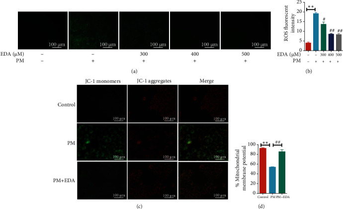
Figure 2.

EDA inhibited PM-induced oxidative stress and mitochondrial dysfunction in HBECs. (a) HBECs were treated with EDA in a dose-dependent manner (300, 400, or 500 μM) and then stimulated with 300 μg/mL PM for 24 h. The ROS level was measured through the fluorescent probe, DCFH-DA, and fluorescence intensity was measured and shown in (b). (c) Mitochondrial membrane potential (ΔΨm) was detected using JC-1 staining, and data are presented as the ratio of red fluorescence (JC-1 aggregates) to green fluorescence (JC-1 monomers) in (d). Values are the mean ± SEM; ∗∗P < 0.01, compared with the control group; #P < 0.05 or ##P < 0.01, compared with the PM group; n = 3. EDA: edaravone; PM: particulate matter; HBECs: human bronchial epithelial cells.
