
Figure 1.
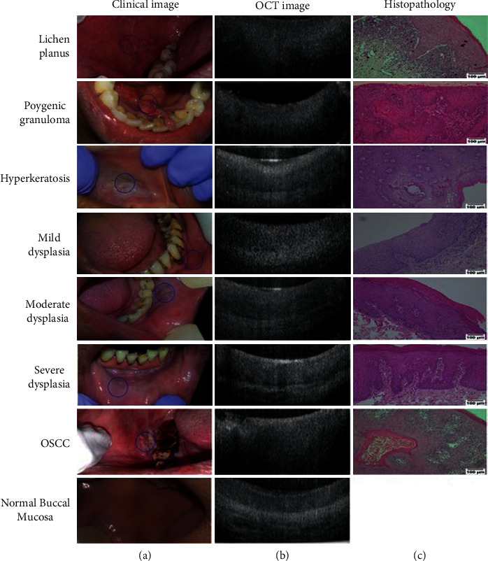
Clinical, OCT, and histological images. Clinical (a) and OCT (b) images were captured from all subjects, and biopsy samples were collected (wherever indicated) and assessed histopathologically (c). Histological images were taken at 100x resolution (scale bar = 100 μm) using Nikon DSFi2 and NIS elements D4 20.0. The nondysplastic lesions shown were histologically diagnosed with lichen planus, pyogenic granuloma, and hyperkeratosis. Normal buccal mucosa images were taken from a healthy volunteer without any habit history. Representative images of all dysplastic grades and a buccal oral squamous cell carcinoma (OSCC) are also depicted (image courtesy of https://bit.ly/316d1S1).