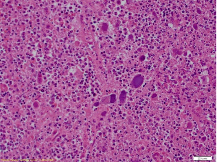
Figure 1. Excision of the lymph node revealed necrotizing granulomatous inflammation (left side of the image) with accompanying residual chronic lymphocytic leukemia/small lymphocytic lymphoma (right side of the image) (100x) (immunohistochemical staining).
Image credit: Henry Ford Health System, Department of Pathology, Jackson, USA.

