. 2020 Aug 24;10(52):31215–31232. doi: 10.1039/d0ra04683c

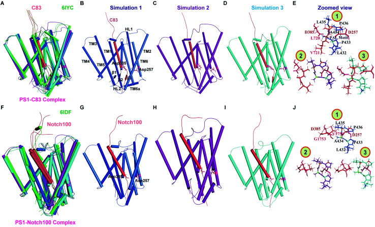
Fig. 4. Comparison of 303 K-simulated PS1 and experimental cryo-EM structures. (A) Superimposed view of simulated and experimental (6IYC) structures of γ-secretase–C83. (B–D) Ensemble-representative simulated PS1–C83 structures. (E) Zoomed-in view of the catalytic residues in the three simulated structures of 6IYC (catalytic and PAL residues are shown as sticks). (F) Superimposed view of simulated and experimental (6IDF) γ-secretase–Notch100. (G–I) Ensemble-representative simulated PS1–Notch100 structures. (J) Zoomed-in view of the catalytic residues in the three simulated structures of 6IDF. Blue, purple and cyan represent simulation 1, 2, and 3 (deep salmon color represents the substrates); the experimental structures are shown in green.

Fig. 4