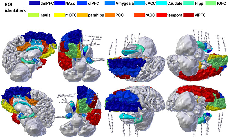
ED Fig. 1. iEEG Recording montage.
Example recording montage from a single participant, with cortical parcellation overlaid. Electrode shanks represented by the grey dotted lines access a broad network covering multiple prefrontal structures, superficial and mesial temporal lobe, and striatum/internal capsule.

