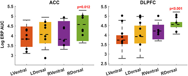
ED Fig. 3. Cortical response to internal capsule stimulation.
Topographic structure of the internal capsule yields differential cortical effects from stimulation at different capsular sites. Before task-linked stimulation, we performed safety/perceptibility testing, where we repeatedly stimulated each potential site with brief 130 Hz pulse trains (see Methods). Each of those trains created an evoked response potential (ERP) in various cortical regions. For each participant, we collected all sEEG channels that were localized to grey matter of DLPFC or ACC. We then quantified the post-train ERP as the sum of the area under its polyphasic curve (AUC). We limited this analysis to channels ipsilateral to the site of stimulation. Each marker represents the mean log(AUC) in one participant. Boxes show the mean and confidence intervals for the ERP AUC from stimulation at each site.
The stimulation sites that were more effective behaviorally produced the largest ERPs in these cognitive-control-associated regions, with right dorsal stimulation having the largest effects. (p-values represent t-test on the regression coefficients of a log-normal GLM, i.e. the same analysis used in main text Fig. 2). In the left hemisphere, dorsal stimulation produced larger responses than ventral stimulation, but this did not reach statistical significance given the small number of trials (5 test trains per participant).
These results are consistent with the known topography of the internal capsule, where fibers that connect DLPFC and ACC to thalamus run in the dorsal-most part of the anterior limb, i.e. in close proximity to our chosen dorsal electrodes.

